

Impacted Canine Tooth: Signs, Assessment and Treatment Options
An impacted canine tooth is a canine that has not erupted into its expected position and remains partly or completely beneath the gum. It does not automatically require the same treatment in every person. Its location, the available space, the health of nearby teeth and the patient’s stage of dental development all influence the plan.
We assess the whole situation before discussing an appliance or operation. In this article, we explain why canine teeth become impacted, the signs that may lead to referral, what imaging can show, and the main management options. We will also describe how an orthodontist, general dentist and oral surgeon may work together when a buried tooth needs to be uncovered and guided into the mouth.
What is an impacted canine tooth?
The canines are the pointed teeth near the corners of the dental arch. The upper permanent canines usually travel a comparatively long eruption path before appearing beside the lateral incisors. When that path is blocked or diverted, a canine may remain in the jaw rather than erupt normally.
An impacted canine tooth may sit towards the palate, towards the lip or in another position. It may be covered by gum alone or by both gum and bone. The baby canine can sometimes remain in place, which can make the missing permanent tooth less obvious.
The British Orthodontic Society’s patient information on impacted canines explains that management depends on factors including the tooth’s position, age, available space and willingness to have fixed braces. Those are the same kinds of considerations we discuss during individual assessment.
Why might a canine fail to erupt?
There is not always one identifiable cause. Eruption is a biological process influenced by the developing tooth, surrounding teeth, available space and jaw anatomy.
Factors that may be relevant include:
- insufficient room in the dental arch
- an unusual eruption path
- a retained baby canine
- a missing or unusually shaped neighbouring lateral incisor
- an extra tooth or other physical obstruction
- previous trauma affecting developing teeth
- a cyst or another local condition
- family patterns of ectopic or impacted teeth
Finding one factor does not explain every case. This is why we avoid planning from a photograph or the appearance of the baby tooth alone.
What signs can suggest an impacted canine?
An impacted canine tooth often causes no pain. It may first be noticed during a routine dental examination or when an expected tooth does not appear. The absence of discomfort does not establish whether the buried tooth is harmless.
Signs that may prompt assessment include:
- one upper canine appearing while the other does not
- a baby canine remaining longer than expected
- a permanent canine erupting high in the gum or away from its usual position
- crowding or loss of space where the canine should emerge
- movement or unusual angulation of a neighbouring tooth
- a swelling in the palate or gum
- an X-ray showing that the canine is not following its expected path
For a child or adolescent, timing is based on dental development rather than a birthday alone. Our article about when a child should have an orthodontic assessment explains why an early visit may lead to monitoring rather than immediate treatment.
Why is assessment important if nothing hurts?
Most concerns are easier to understand before making irreversible decisions about the baby canine or neighbouring teeth. A buried canine may remain stable, but in some positions it can affect the root of a nearby incisor or develop another problem. The level of risk is individual.
Assessment aims to answer four practical questions:
- Where is the permanent canine?
- Is its path affecting another tooth?
- Is there enough space for it in the arch?
- What are the reasonable options at this stage?
The answer may be review, an interceptive step or a longer treatment plan. We do not imply that every impacted tooth needs to be pulled into place.
What happens during the orthodontic assessment?
We begin with the history. We ask about the timing of tooth loss and eruption, previous dental trauma, family patterns and any symptoms. We then examine the teeth, bite, space and the contour of the gum and palate.
Records are selected according to the clinical question. They may include photographs, a digital scan and radiographs. A two-dimensional X-ray can show the general position, while three-dimensional imaging may be considered in selected cases when the relationship to nearby roots cannot be understood adequately another way. Imaging should be justified by the information it is expected to add.
| Part of the assessment | What it helps us understand |
| Dental history | Eruption timing, trauma and earlier treatment |
| Clinical examination | Space, retained teeth, bite and palpable position |
| Photographs or scan | Arch shape and available room |
| Radiographs | Tooth position, development and nearby structures |
| Growth and age assessment | Which options remain realistic and when to act |
After reviewing the records, we explain what is known, what remains uncertain and which options are proportionate.
Can removing the baby canine help?
In selected developing cases, removing a retained baby canine may improve the chance that the permanent canine changes its path. This is not a universal solution, and it does not guarantee spontaneous eruption.
The decision depends on the permanent tooth’s position, development, space and the condition of adjacent teeth. Follow-up is needed because an interceptive step only makes sense if its effect is reviewed. If the canine does not improve its path, another plan may be discussed.
No parent should interpret this as a reason to arrange extraction without orthodontic assessment. A useful plan identifies why the tooth is being removed, what change is expected and when progress will be checked.
What are the treatment options?
The options range from observation to coordinated surgical and orthodontic care. The appropriate route is the one that fits the diagnosis and the patient’s priorities.
| Option | When it may be considered | Important limitation |
| Monitor the tooth | The position is acceptable to observe, and no active problem is present | Requires planned review and sometimes repeat imaging |
| Remove a retained baby canine | A developing permanent canine may have a reasonable eruption path | Does not guarantee that the permanent tooth will erupt |
| Create space orthodontically | The arch lacks room for the canine | Space alone may not correct the tooth’s direction |
| Surgical exposure and orthodontic traction | The canine can reasonably be guided into the arch | Involves surgery, fixed appliances and an extended sequence |
| Remove the impacted canine | Bringing it into place is unsuitable or risks outweigh benefits | Leaves a restorative or space-management decision |
| Accept another tooth arrangement | The bite and appearance can be managed without the canine in its usual position | Suitability depends on the full dental relationship |
We discuss benefits, burdens, uncertainties and alternatives rather than presenting one method as the answer for everyone.
How do exposure and orthodontic traction work?
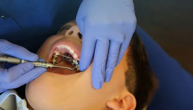
When exposure and traction are chosen, space is usually created or confirmed first. An oral surgeon then uncovers the impacted canine and may attach a small button or chain. The orthodontic appliance applies light, planned force to guide the tooth gradually.
The sequence can include:
- Preparing the arch and making sufficient space.
- Surgical exposure by the appropriate clinician.
- Healing and attachment management according to the agreed technique.
- Gradual orthodontic traction.
- Positioning the canine and coordinating the bite.
- Retention after active movement.
Movement is not immediate. Distance, direction, bone, root shape, ankylosis and the response of surrounding tissues can all affect progress. We explain these uncertainties before treatment begins.
What are the risks and limitations?
Material risks depend on the plan. They may include discomfort, gum changes, appliance breakage, root shortening, damage already present in adjacent teeth, failure of the canine to move, or the need to reconsider treatment. Surgery has its own risks, which the surgeon explains.
An impacted canine tooth can occasionally be ankylosed, meaning it is fused to surrounding bone and does not respond normally to orthodontic force. This cannot always be predicted with certainty at the outset. Treatment planning therefore includes alternatives if movement does not proceed as expected.
Oral hygiene matters throughout. Fixed braces and surgical attachments create extra areas to clean, while inflamed gums can complicate care. The general dentist continues routine prevention and treatment during the orthodontic process.
How long does treatment take?
There is no reliable single duration for every impacted canine tooth. Treatment time depends on how far the tooth must travel, its angle, the space required, age and biology, the appliance plan and whether other bite corrections are being made.
A written plan should describe the expected stages without promising an exact result or finish date. Missed appointments, breakages and poor elastic or hygiene routines can also affect timing.
What should parents and adults ask at the consultation?
A useful discussion should leave you clear about the diagnosis and the choices. Consider asking:
- Where is the canine in relation to nearby roots?
- Is there evidence of damage now?
- Is monitoring reasonable, and what would change that decision?
- Does space need to be created before another step?
- Who would perform any surgical exposure?
- What appliances are likely to be used?
- What happens if the tooth does not move?
- What care will my general dentist provide during treatment?
- What is included in the written estimate?
These questions make uncertainty visible. They also help distinguish a treatment objective from the tools used to pursue it.
Arrange a specialist orthodontic assessment
Michael Woods Orthodontics has two specialist orthodontists and has practised from one site since 1988. Professor Michael Woods’ stated clinical focus includes early interceptive orthodontics and complex care. We state those facts so you can understand the clinical setting, not as a promise about an individual outcome.
If a dentist has identified an impacted canine tooth, bring the referral and existing X-rays where available. Call us on (03) 9509 7496 or request an appointment. We are on Dandenong Road, on the Malvern/Armadale border.
Frequently asked questions
1. Does every impacted canine tooth need surgery?
No. Monitoring, interceptive extraction, surgical exposure, removal or another space-management plan may be considered depending on the case.
2. Can an adult impacted canine be treated?
Sometimes. Position, supporting tissues, ankylosis risk, available space and the condition of nearby teeth influence suitability. Age alone does not answer the question.
3. Will removing the baby canine solve the problem?
It may help in selected developing cases, but it cannot guarantee eruption. The permanent canine’s position and follow-up findings matter.
4. Can clear aligners bring down an impacted canine?
Appliance choice follows the required movement. An impacted canine that needs surgical exposure and traction often requires fixed or auxiliary components. We assess the full sequence before discussing a specific appliance.


Leave a Reply